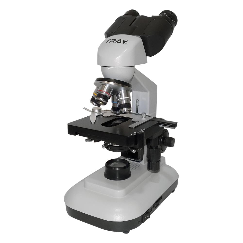
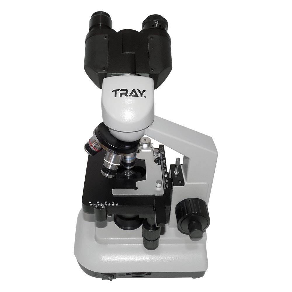
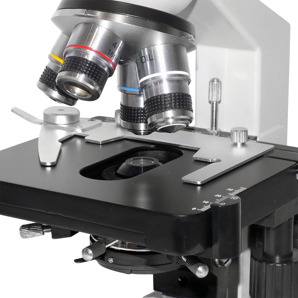
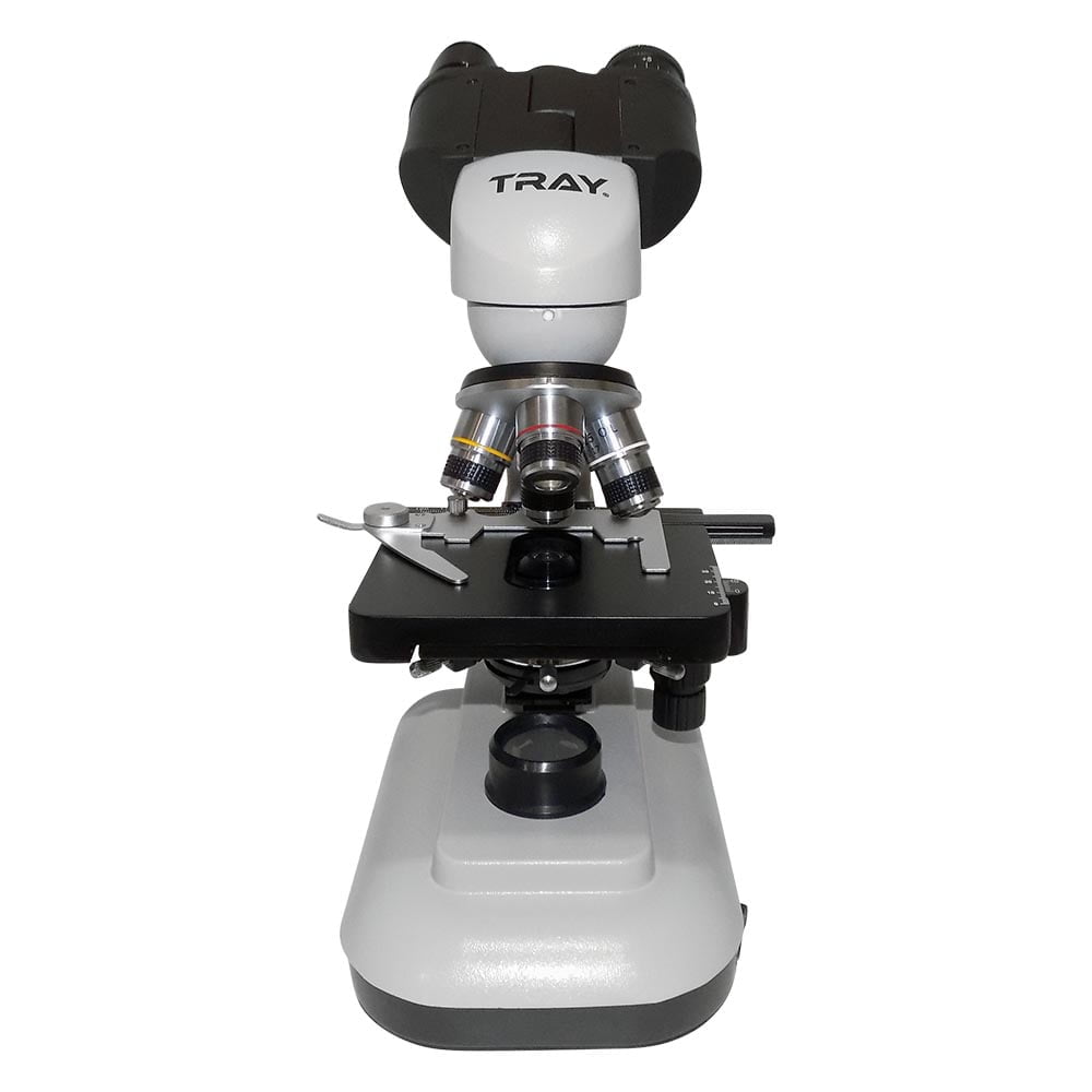
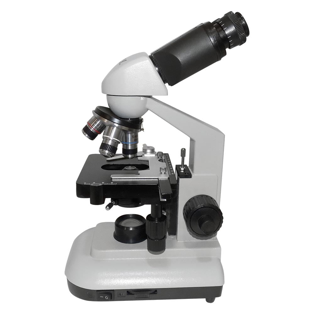
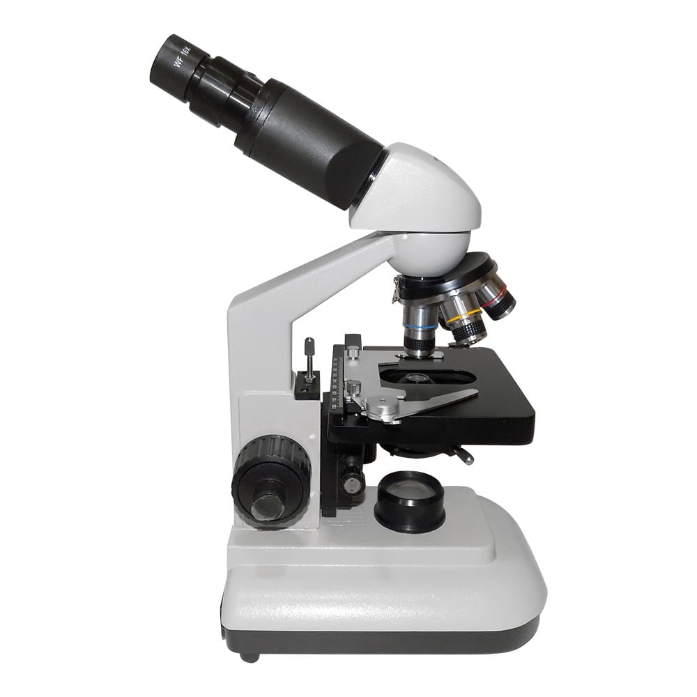
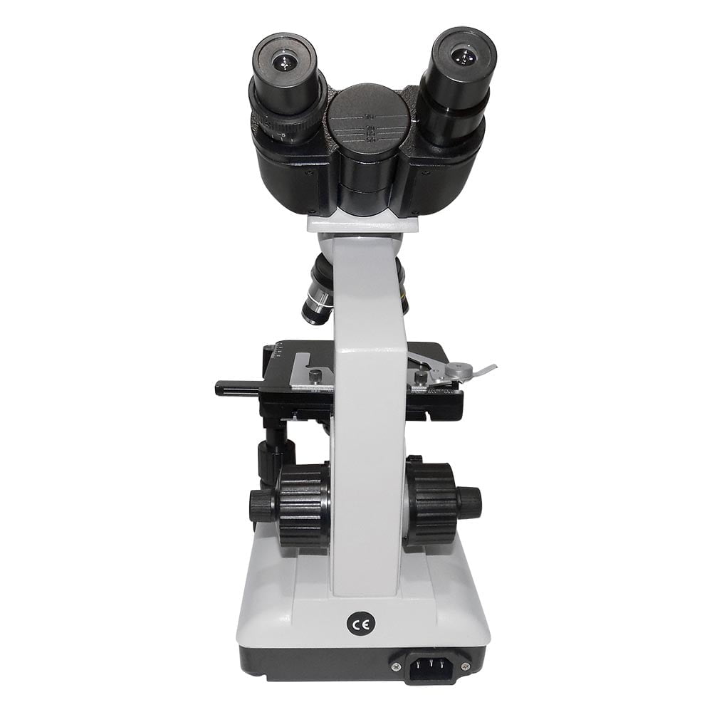
Biological microscopes are mostly used for observing and testing biological research in research laboratories, universities and secondary schools. It is also used for routine testing, clinical testing, and educational and training demonstration in medical and healthcare facilities and laboratories. The microscope magnifies from 40 times to 1000 times. Additionally, with the WF16 eyepiece, magnification can go from 640 times to 1600 times.
Microscope packaging consists of foam and cardboard. The main components are in plastic bags. Lenses should be carefully removed from the bag and mounted. Likewise, we must carefully attach the eyepiece lens. The objective and eyepiece lenses should not be touched by hand, and parts of the microscope should not be disassembled or assembled.
The optical imaging and illumination principle of the microscope is shown as a diagram.
The imaging system consists of objective, prism and eyepiece. The lens primarily magnifies the sample and the light rays are refracted by the prism up to 45 degrees and captures the image in the ocular imaging plane, then it is observed with the eyes by secondarily magnifying it. Total magnification is determined by the product of the magnification of the objective and the eyepiece.
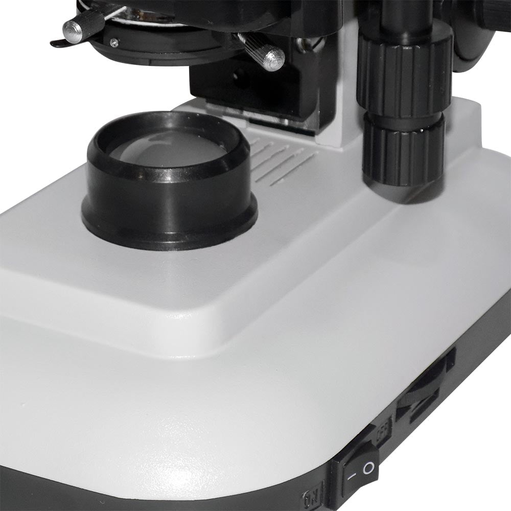
The lighting system consists of lamp, collector, diaphragm and capacitor. Light rays from the lamp pass the collector and illuminate the diaphragm, then are combined by the capacitor. This system can illuminate the observed sample on stage for visual observation. You can illuminate it with a reflector to replace the lamp.
Eyepieces
| Magnification | Diameter of view-field(mm) |
| Wide field eyepiece | WF10X | 16 |
| Wide field eyepiece | WF16X | 11 |
Objectives
| Magnification | Numerical aperture | Working distance | Remark |
| Achromatic objectives | 4X | 0.10 | 37.5 | |
| 10X | 0.25 | 7.613 | |
| 40X(s) | 0.65 | 0.632 | Spring |
| 100X(S,OIL) | 1.25 | | Spring, oil |


